The Top 5 Uses of Adhesive Microscope Slides in Today’s Laboratories
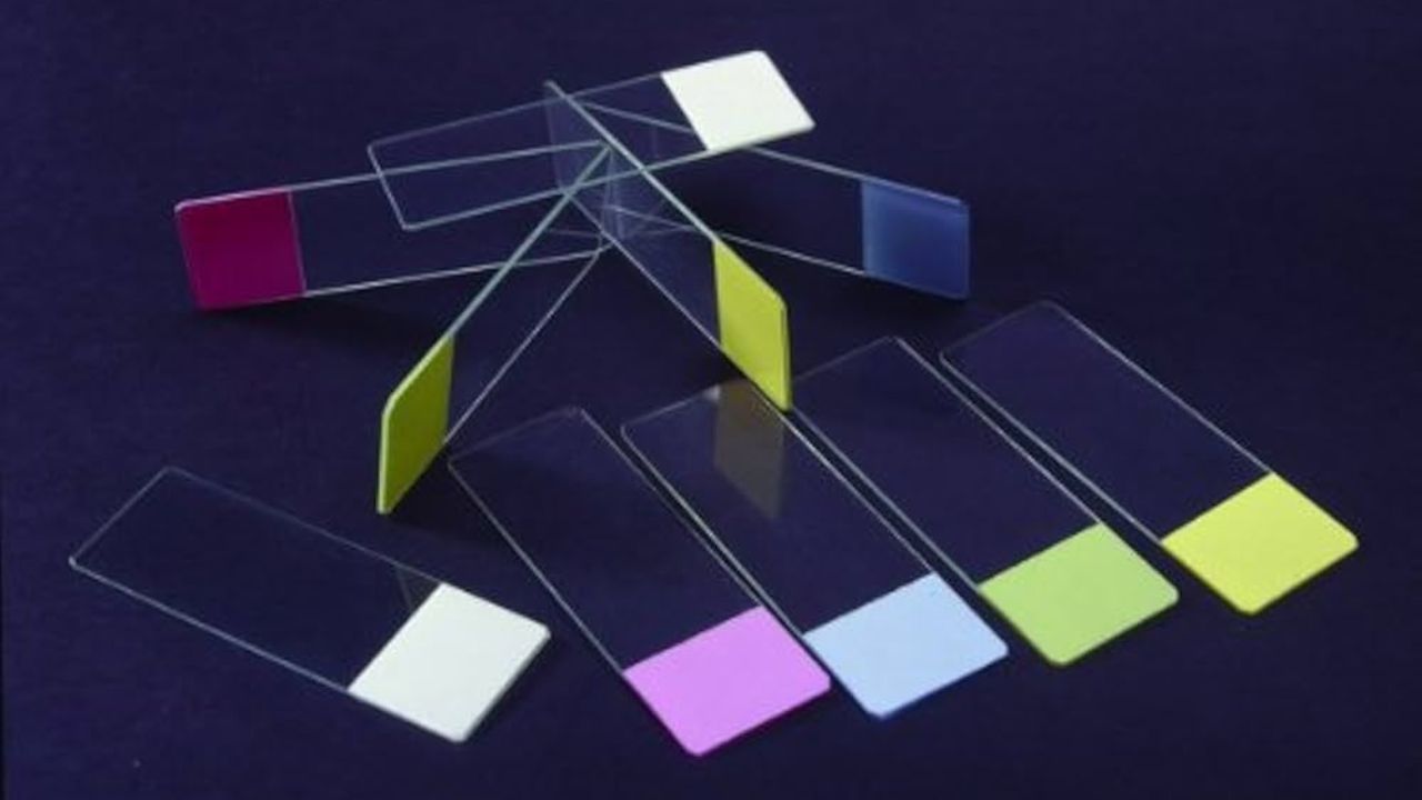
Adhesive microscope slides have become crucial in modern labs. Especially those involved in advanced research and healthcare. These slides have a special coating, which can be polysilane or silane, that improves the bonding of specimens. This form of binding is needed to hold biological samples during all stages of processing and analysis. Because laboratories are using more advanced methods, microscope slide adhesive is being used for many more applications. These laboratory microscope slides play an important role in today’s labs for the following five reasons.
Immunohistochemistry (IHC)
Specific antigens in tissue sections are detected using immunohistochemistry and antibodies. Those are marked with visible substances. Many use this technique in cancer diagnostics, biomarker studies, and for developing new drugs. Adhesive microscope slides help by holding thin tissue sections together as the tissue is processed several times, including heating, using enzymes, and washing. When there is not enough adhesive on the surface, samples may come off, degrade, or shift, resulting in inaccurate outcomes. Tissue slides coated with silane or charged positively are the best choice for IHC since they ensure the tissue stays in place under all circumstances. By using these techniques, the shape of the tissue and where the antigens are found do not change during staining.
In Situ Hybridization (ISH)
This technique in molecular biology is used to detect and locate particular nucleic acid sequences in cells and tissues that have been preserved. People use it in genetics, early screening for babies, and testing for infectious diseases. During ISH procedures, samples need to be stable since they are treated with chemicals and exposed to numerous hybridization conditions. Microscope slides with polysine or similar agents ensure that cells and tissues with nucleic acids do not detach during the process. A strong adhesion between the probe and the sample is necessary to avoid losing the sample, which could reduce the clarity and specificity of the signals. Adhesive slides are used because they help ensure that specimens are reliably positioned during in situ hybridization.
Cytology and Cell Smear Analysis
To perform cytological analysis, cells are examined to look for any abnormalities, infections, or cancer. Doctors may use routine Pap smears, check urine for changes, and take fine needle aspirates. Here, cells are applied to a microscope slide, fixed, and stained. Adhesive slides are necessary for cytology. Because they prevent cells from being lost when preparing the sample. Sometimes, scientists prefer silane or hydrophilic-coated slides, as the surface supports a variety of cells, including epithelial cells, lymphocytes, and neoplastic cells. Not altering the structure of cells is a big benefit for getting accurate views under the microscope.
Cell Culture Monitoring and Research
Researchers in cell culture studies use adhesive microscope slides to keep an eye on cell growth, their shape, and what they do in the laboratory. Sometimes, cells are grown straight on a slide to allow for easy observation by the microscope or for future staining. Stem cells, neurons, and fibroblasts attach and spread well on hydrophilic and silane-coated slides. They are used to ensure that cells grow in the same way every time in drug testing, developmental biology, and regenerative medicine research. Using the same platform for culture and analysis allows for easier and safer work in the laboratory.
Tissue Sectioning and Histological Analysis
Histology studies the small details of tissues, which are usually observed using sections from FFPE tissue that have been treated with formalin and paraffin. Once the tissue blocks are thinly cut, the pieces go on slides, are stained, and then looked at under a microscope. Adhesive slides are needed because the tissue sections can come loose at some points during processing. Silane-treated and positively charged slides ensure the sections are well attached and stay intact after being treated at high temperatures. Because of their stability, histologists and pathologists can do in-depth examinations of tissues, aiding in the identification of diseases such as cancer, autoimmune diseases, and conditions that worsen over time.
Conclusion
Using adhesive microscope slides has made it much easier to prepare and study samples in various areas of science and medicine. The enhanced ability to stick makes them reliable, always consistent, and precise, which is necessary in difficult laboratory situations. Using specialized slides allows for histopathology, cytology, genetic analysis, and cell culture, keeping samples intact and improving the accuracy of the analysis. Many laboratories find that adhesive microscope slides are a necessary tool for them to rely on each day. As scientific methods develop, the need for these excellent slides will only increase.